Imaging
We make available to the research community microscope and imaging systems capable of multiple modes of imaging of both live and fixed biological samples, including:
- Confocal microscopy (resonant, point scanning and spinning disc)
- Super Resolution microscopy (STED)
- Widefield fluorescence microscopy
- Light sheet microscopy
- Automatic plate/slide imagers
- Kinetic imaging system
- Image analysis software
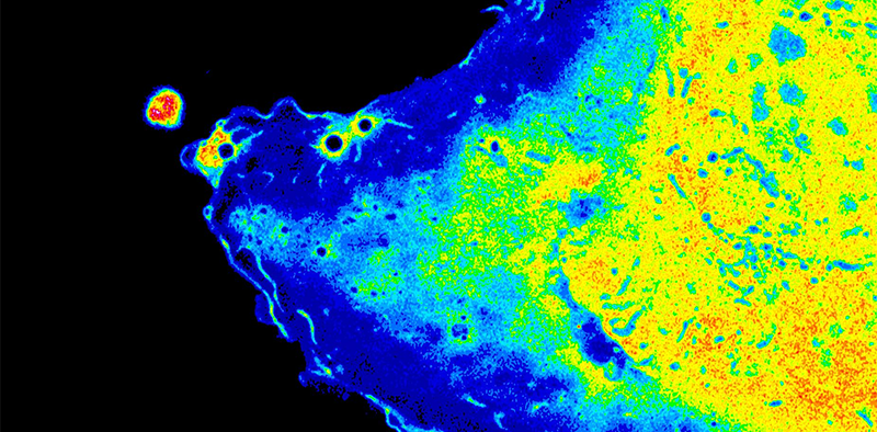
Live Airyscan confocal image of migrating epithelial cell expressing the genetically encoded hydrogen peroxide sensitive fluorescent protein HyPer3 (Thiagarajah Lab)
Cell function and characterization
We provide the infrastructure, expertise, and training for:
- Multi-color flow cytometry
- Multi-color cell sorter
- SeaHorse Analyzer
- MERFISH
- Single Cell Transcriptomics
Gaining access
Our equipment is open for use to all Boston Children’s Hospital researchers. See current pricing, request training and access equipment scheduling, by clicking the "Request Services" button below. Please login with your existing iLAB account or create a new account, and then search for Cell Function and Imaging Core (Harvard Digestive Diseases Center).